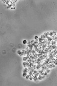


Alnus glutinosa
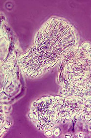
Morella pensylvanica
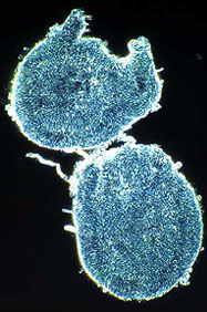
Coriaria plumosa
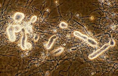
Hyphal Clusters isolated from root nodules (Photos by D. Benson)
Hyphae measure about 0.5 to 1.5 μm wide. The cell walls appear two layered (Baker, 1980; Lalonde, 1979; Lancelle, 1985; Horriere, 1983), with cross-walls initiating from the base layer. Lipid layers are sometimes seen outside the wall that probably correspond the lipid layers so prominent on vesicles (Harriot and Benson, 1991; Horriere, 1983; Newcomb, 1979; Newcomb, 1987).
The cytoplasm of hyphae contain rosette-shaped granules presumed to be glycogen as well as lipid bodies (Benson, 1979; Lancelle, 1985). Frequently seen are cytoplasmic tubules that are circular in cross section, and average 45 nm in diameter. These structures underlay the cell membrane both at the cell septum and the outside wall (Lancelle, 1985). Aerial hyphae are not produced on solid media.
The spores are "sporangiospores" borne in "multilocular" sporangia that are formed either terminally, or in an intercalary position on the hyphae (below; more on spores).

All Frankia strains tested differentiate "vesicles" in N-deficient culture, and often in symbiosis. Vesicles are lipid-encapsulated, roughly spherical cellular structures measuring between 2 to 6 um in diameter, attached to hyphae by a short vesicle stalk that is also encapsulated; they are commonly made in response to nitrogen deprivation. Vesicles contain nitrogenase and ancillary enzymes. (More on vesicles)