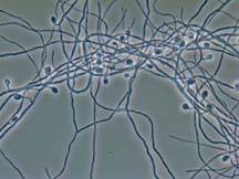
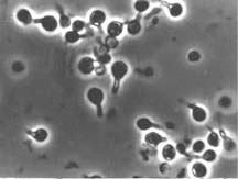
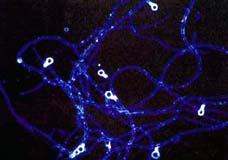
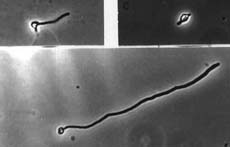
All Frankia strains tested make "vesicles" in N-deficient culture, and often in symbiosis. Vesicles are lipid-encapsulated, roughly spherical cellular structures that measure between 2 to 6 μm in diameter. They are attached to hyphae by a short vesicle stalk that is also encapsulated. Vesicles contain nitrogenase and ancillary enzymes.
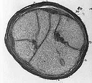
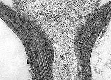
Vesicle Envelopes
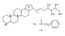
Vesicles have a laminated lipid envelope (Lamont et al., 1988; Torrey and Callaham, 1982). The number of laminae is correlated with the pO2 of the surrounding medium (Parsons et al, 1987). The layers are extracted during fixation for TEM, leaving a "void area" (Lalonde et al, 1976) but they can be visualized by permanganate fixation (Harriott et al., 1991). Each layer is about 4 nm thick. Isolated vesicle envelopes are comprised primarily of hopanoid lipids (Harriott et al, 1991; Berry et al., 1993).
Bacteriohopanes play a role in membrane stability and fluidity (Demel et al, 1976; Kannenberg et al., 1980). In Frankia vesicles, they may limit O2 diffusion into the O2 labile nitrogenase.
As vesicles senesce, septa become irregular, nitrogenase activity ceases, and the cytoplasm disappears. Some vesicles can, however, 'germinate' and produce hyphae in culture (Schultz and Benson, 1989).