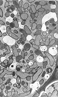
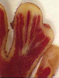
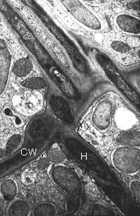
In the plant cell Frankia hyphae and vesicles are encapsulated with plant cell wall-like material and remain outside the host plasmalemma. The capsule contains pectin, cellulose and hemicellulose (xylans) (Lalonde, 1975). The capsule has been described as "...a thin internal tubular plant cell wall" (Berg, 1990). In Ceanothus the capsule has a very high concentration of polygalacturonans and appears distinct from the cell wall (Liu et al, 1991). The infected zone is often delineated by lignified cell walls.
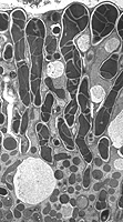
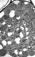
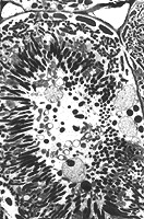
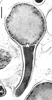
Hyphae pass through the middle lamella of cell walls and host cytoplasm. "Colonizing hyphae" penetrate cell walls and "proliferating hyphae" branch radially in host cells (Newcomb, 1987). As Frankia mature, symbiotic vesicles take on a variety of appearances depending on the plant. Alnus and Elaeagnus have large spherical, mulitseptate vesicles, while members of the Rosaceae and Ceanothus have non-septate elliptical vesicles. Datisca and Coriaria have hyphal vesicles that are interdigitated with host mitochondria, presumably to maintain low pO2 levels in the vicinity of nitrogenase. Myrica, and Comptonia have club-shaped hyphal swellings while Casuarina has an entirely filamentous structure (Newcomb, 1987). Vesicle morphology in symbiosis is controlled by the host plant since all isolated frankiae produce spherical vesicles in culture. Freeze-fracture electron microscopy has revealed that symbiotic vesicles contain the multilayered envelope like that made in culture (Abeysekera, 1990; Lalonde, 1976; Newcomb, 1987)