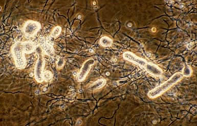
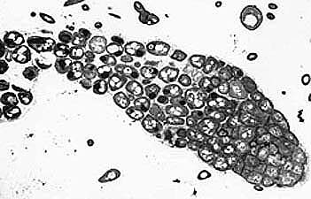
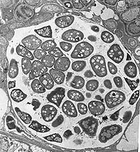
Mulitlocular sporangia are produced in culture by all Frankia strains. Sporulation also occurs in some nodules, especially in Myrica and Alnus (Schwintzer, 1990; Torrey, 1987). Sporangia develop as terminal or intercalary structures on hyphae (Newcomb, 1979). Segmentation within the enlarging sporangia produces a multilocular sporangium containing many spores. Electron microscopy shows the developing spores with electron translucent nucleoid regions with dispersed fibrils and numerous lipid droplets. Like the hyphae, the external walls often show the residue of a laminate envelope (Lancelle, 1985). The mature spores show evenly dispersed cytoplasm but tubules are not present in the developed spores (Lancelle, 1985).
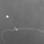
Sporangia develop by hyphal thickening, then segmentation by septa originating from the inner layer of a double-layered sporangial cell wall (Horriere, 1983; Lancelle, 1985; Baker, 1980; Newcomb, 1979; Horriere, 1983). The result is that older, more loosely packed spores, are distal to younger, more densely packed, and developing spores. The intersporal matrix appears to originate from the inner layer of the cell wall. Younger spores are more irregularly shaped, and older spores are spherical to ovoid, and measure about 1-5 μm in size. These sporangiospores are nonmotile, unlike those found in the multilocular sporangia from Geodermatophilus or Dermatophilus. Sporangia contain a few to several hundred spores depending on the age, and nutrition of particular strains.